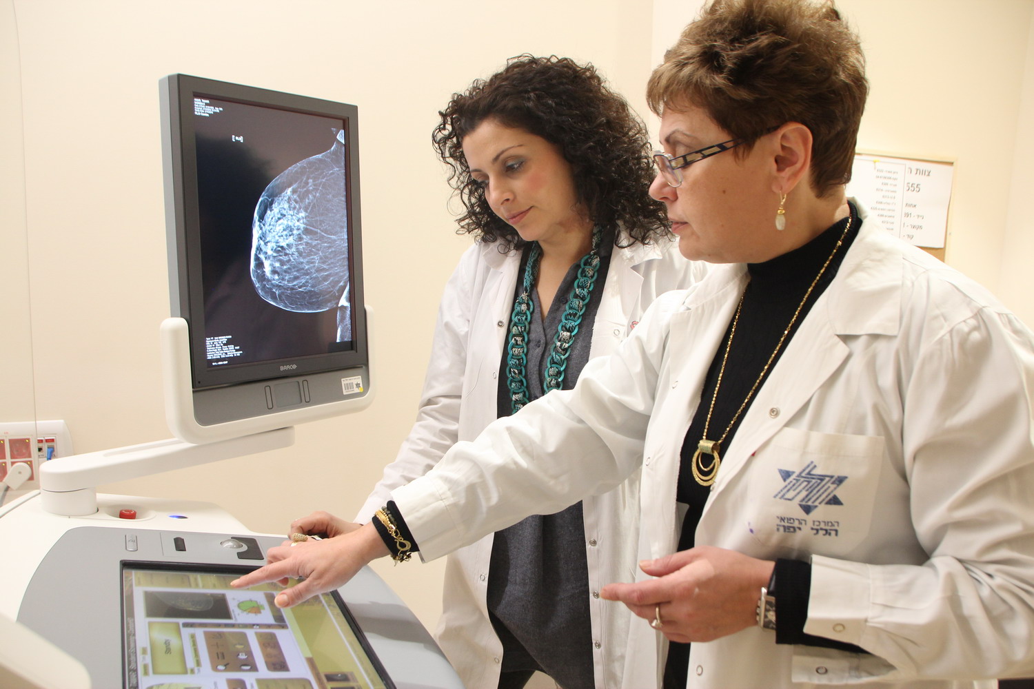
Breast cancer is the most common form of cancer in women. It is estimated that 1.7 million new cases of breast cancer are discovered annually worldwide. In Israel, as well, it is the most common disease among women, and each year over 5,000 new cases are diagnosed. October is Breast Cancer Awareness Month. Director of the Imaging Institute, Dr. Natalia Kochnovski and Head Radiographer at the Institute, Tagrid Younis, explain the importance of early detection and the means currently available to diagnose and detect breast cancer.
Facts and figures
About three years ago, Hillel Yaffe opened its Breast Imaging Institute. The staff, made up solely of women, makes sure to provide patients with personal care, guidance and close follow-up. “Every woman who comes to us is fearful and concerned, especially when a tumor is suspected. The fact that we are all women helps the patients feel more comfortable, allows them to open up and share their feelings. When a woman is suspected of having a malignant tumor, we immediately complete the investigation, including performing a biopsy, and don't leave her in a state of anxiety and uncertainty, which is difficult to cope with in and of itself,” said Tagrid.
Over 7,500 mammograms and ultrasounds have already been performed on thousands of women, including 1,200 biopsies. The interesting statistic is that approximately 25% of the biopsies found malignant growths. “This figure should sound an alarm for all women, particularly those over the age of 40 and those with a high risk of breast cancer. There is no disputing that early detection does, in fact, save lives and increases the odds of recovery and return to routine,” said Dr. Kochnovski. “Therefore, all women approaching 50 or anyone who feels a new lump, experiences discharge from a nipple or unusual pain - should go have a manual exam performed by a breast surgeon and continued investigation, if necessary.”

Right to Left: Dr. Natalia Kochnovski and Tagrid Younis at Hillel Yaffe's Breast Imaging Institute
Types of exam
Today, thanks to innovative and advanced technology, there are several different exams to diagnose and detect both benign and malignant tumors:
Screening mammogram - breast X-ray when there are no symptoms. The only test approved as screening for early detection of breast cancer. According to the Ministry of Health's guidelines, screening once every two years is recommended for all women over the age of 50, without clinical findings or a background of breast or ovarian cancer in the family. "However, women may begin having mammograms every year or two, starting at the age of 40– according to the guidelines of the physician interpreting the image. Women who are at a higher risk of having malignant tumors or who have breasts that are very dense, where it can be difficult to detect malignant tumors, may be sent for a mammogram annually, even if they are under 50,” said Dr. Kochnovski. It is important to note that during pregnancy and when breastfeeding, screening mammograms are not performed due to higher sensitivity to the effects of radiation, lower efficacy due to the density of the breast and benign findings that are common during pregnancy that may appear to be malignant. The exam may be performed about three months after pregnancy and/or breastfeeding.
Diagnostic mammography - a breast X-ray of women who have symptoms or indications of breast disease, patients who have had a screening mammogram with findings that called for continued investigation or patients with mammogram findings that require continued follow up.
Digital mammography, including tomosynthesis, for the purpose of screening and diagnosis - “We work with the latest digital mammography device, which enables precise diagnosis of breast cancer, in addition to standard mammography,” said Tagrid. “The new technology was developed in recent years in order to reduce failures in diagnosis of breast cancer among women with dense breasts. It is three-dimensional mammography, during which several images of the breast at various angles are produced, at thin cross-sectional “slices,” which enable the texture of the breast to be seen clearly and more precisely, to identify lesions in the breast, without overlap of adjacent breast tissue and to reconstruct the pictures into regular breast images. This significantly reduces the amount of radiation to which the woman is exposed in the mammography exam,” stated Tagrid.
Ultrasound - a non-radioactive test that is performed to complete the investigation of a suspicious finding, for absolute distinction between cysts (lumps that contain fluid and are benign) and solid lumps that are not filled with fluid and between benign lumps and tumors, to follow-up on known findings, to assess inflammatory processes and as a tool for any type of invasive procedure.
Mammography- or ultrasound-guided biopsy - when an unclear or suspicious finding is found, the next step in the investigation is a biopsy. The biopsy is performed according to the level of suspicion, when it is necessary to verify or rule out the existence of a malignant tumor.
MRI – the most sensitive exam for detecting breast cancer, without X-ray radiation. It is generally performed on women who are at high risk or are genetic carriers, to follow-up on findings from previous tests, continued investigation of mammography findings or for women with silicone implants.
The ball is in your court
As we have already stated, early detection of breast cancer saves lives and, therefore, the ball is in your court. The following are some recommendations:
-
If you are over 50 - it is recommended that you a screening mammogram.
-
If you are over 40 - it is recommended that you consult with a breast surgeon to check your risk level and, if necessary, to begin investigation through a mammogram.
-
If you feel a new lump in your breast, if the skin on your breast changes color, you experience discharge from your nipple or anything that looks or feels different - go to be examined by a breast surgeon.
As stated, screening exams are not performed during pregnancy and when breastfeeding, as the structure of the breast changes. However, if you are pregnant or breastfeeding and there is a suspected abnormal finding, you should go be examined by a breast surgeon who will refer you for a mammogram if necessary.
Don’t hesitate - go get tested!
To schedule an appointment at the Hillel Yaffe Breast Imaging Institute, call 04-7748322.