Nuclear medicine is a discipline that uses radioactive substances to diagnose and treat through hybrid imaging.
The advantage of nuclear scanning compared to other imaging methods is that it demonstrates changes in the function of the examined organ and not only structural (anatomical) changes. Therefore, at times nuclear scans can identify certain diseases earlier than other imaging methods.
The scan is performed at the same time or following injection of a radioactive substance. The waiting time between the injection and scan with the camera differs by scan and the substance injected.
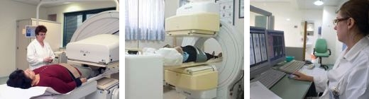
The institute has two new tomographic (3D imaging) cameras that include SPECT-CT and two ergometry rooms for stress testing.
Tests performed at the institute:
-
Bone scans (planar, dynamic, SPECT-CT)
-
Scanning with DPD to check cardiac amyloidosis
-
MIBI or SPECT thallium cardiac scans
-
Brain perfusion SPECT scan
-
Gastric emptying scintigraphy
-
Parathyroid MIBI (SPECT-CT) scans
-
DMSA/DTPA kidney scans
-
Thyroid scans
-
UPTAKE
-
Treatment of hyperthyroidism with iodine
-
Adrenal gland scans o Liver and spleen scan
-
MUGA heart scan
-
Pulmonary perfusion scan/SPECT-CT perfusion
-
Gallium scans (SPECT-CT) to investigate an inflammatory process/sarcoidosis
-
Salivary gland scans
-
Tear duct scans
-
Scans to investigate Meckel's diverticulum
-
Scans with tagged red blood cells (RBC) to detect hepatic hemangiomas and identify acute bleeding in the digestive system
-
Lymphoscintigraphy of the lymph nodes and lymph ducts - to identify a sentinel lymph node and investigate edema in the limbs (lymphedema) | 


.jpg?BannerID=39 "youtube channel")