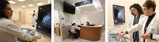
The Breast Imaging Unit at Hillel Yaffe Medical Center is engaged in early detection of breast cancer, through the use of several forms of imaging - some of the most advanced and innovative in the field, depending on what is necessary: digital mammography, including tomosynthesis for screening and diagnosis, ultrasound, ultrasound-guided biopsy and mammography with tomosynthesis, marking prior to surgery, placement of clips prior to chemotherapy and more.
The unit uses the most advanced and innovative equipment, some of which is only available at very few large centers in Israel (such as mammography with tomosynthesis and tomosynthesis-guided biopsy). In some cases, it is the only center in Israel with the equipment (automatic robotic ultrasound to scan the entire area of the breast).
Services provided by the unit
Digital mammography, including tomosynthesis, for screening and diagnosis - X-ray of the breast using the tomosynthesis method - a 3D mammogram, during which several images of the breast are taken from different angles, in thin 1 mm cross-sections, which make it possible to see the texture of the breast more clearly and accurately, to identify lesions without overlap of nearby breast tissue, as occurs in standard two-dimensional mammography. Another advantage offered with this method is the possibility to reconstruct the images as standard breast images, without the additional radiation to which women are exposed during a mammogram. The new technology is particularly good for women with dense breast tissue (more gland tissue than fatty tissue), which is especially typical of young women and menopausal women receiving hormone therapy.
Screening mammogram - breast X-ray of women who do not have symptoms. Mammography is the only test approved as screening for early detection of breast cancer. According to the Ministry of Health guidelines, all women between the ages of 50-74 are entitled to have a mammogram once every two years as part of the national screening program. However, it is recommended that women begin having mammograms every year or two, beginning when they are 40, according to the guidelines of the physician interpreting the image. Mammograms can continue being performed every year or two even over the age of 74 - there is no upper age limit at which point mammograms are no longer effective.
Diagnostic mammography - a breast X-ray of women who have symptoms or indications of breast disease, women who have had a screening mammogram with findings that called for continued investigation or women with mammogram findings that require continued follow up.
Contrast-enhanced mammography - A mammogram that uses a contrast agent. This test is performed in special cases, in order to receive a more precise diagnostic result.
Ultrasound - A test without radiation using the most innovative device, which supplements a mammogram or to investigate a finding felt in the breast. Ultrasound makes it possible to definitively distinguish between cysts (benign lumps that contain fluid) and solid lumps that do not contain fluid. Moreover, it is also possible to distinguish between benign and malignant tumors.
Automatic robotic ultrasound for screening the entire area of the breast - This test is performed using an innovative device, the only one of its kind in Israel, to screen the entire area of the breast. A test using the robotic device for automatic ultrasound screening of the breast resolves the issue of dense breast tissue and enables a supplemental test to the ultrasound screening test recommended every year or two. Systematic and uniform screening of the entire area of the breast is done once every period and can be compared, allowing for follow-up of findings.
Mammography- or ultrasound-guided biopsy (including tomosynthesis guidance) - If an unclear finding is found, the next step will be investigation through biopsy. Biopsy is used in cases where there is a significant probability that there is normal texture and in cases when they suspect that a finding is cancerous and need unequivocal results in order to plan surgery, its course and/or presurgical chemotherapy. The options available in this field of investigation are open biopsies (operating room) and closed biopsies. The institute only performs closed biopsies.
MRI - An MRI is the most sensitive test in identifying breast cancer. The test does not involve exposure to X-rays. During the test, a contrast agent - gadolinium - is injected into the vein of the patient, who is lays on her stomach throughout the test, approximately 25 minutes. Indications for MRIs: genetic carriers, women with a high risk, follow-up on findings from earlier tests, patients diagnosed with carcinoma for continued investigation of the mammography or sonar findings, women with silicone implants.
Breast biopsy under MRI guidance - Women who have had a breast MRI which showed a suspicious finding that requires a biopsy may undergo a breast biopsy under MRI guidance at Hillel Yaffe Medical Center. This is done under local anesthesia and is not painful. Click here for more information >> | 


.jpg?BannerID=39 "youtube channel")